
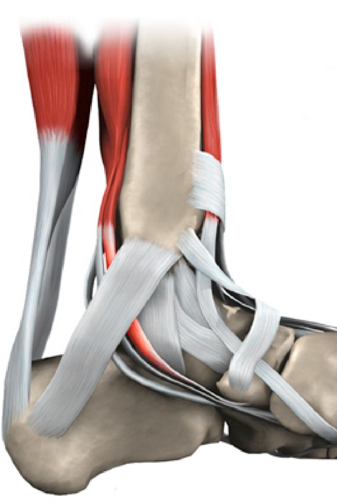
The tibialis posterior (TP) tendon is one of the major stabilising structures in the foot. It runs behind the bump on the inside of the ankle (the medial malleolus) and inserts into one of the bones of the instep (navicular). The main functions of the tendon are to support the arch and keep the foot turned inwards when walking. The TP can become damaged by wear and tear or acute trauma.
Initially, pain is felt along the length of the tendon behind the inside of the ankle. With time deformity becomes apparent and the foot flattens and turns outwards. Pain may develop on the outside of the ankle and if the deformity continues to worsen over time the joints in the hind foot become affected and can become arthritic. The surgical treatment is complex and depends on the location and severity of damage.
Non-Operative Management
In the early or mild stages of TP tendon dysfunction, simple painkillers, orthotics, and physiotherapy are used. Sometimes this is not sufficient and ankle bracing or the use of a custom moulded splint is required. If these non-operative methods prove inadequate to control symptoms or the problem progresses, surgery may be helpful.
Operative Management
In most cases the tendon itself is not able to be repaired and needs to be strengthened by using a near-by tendon which functions to flex the toes and this is transferred from the foot. Other tendons help to carry out this function so loss of this tendon rarely results in problems.
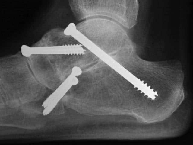
To improve the biomechanics of the tendon transfer the heel bone is moved towards the inside of the foot (calcaneal osteotomy) and held with screw fixation. In addition, a plug may is inserted into the outer portion of the foot to assist in supporting the arch. If required the plug and/or screws can be removed in a second operation when they have served their purpose and the tendon is healed. Additional procedures such as synthetic tissue augments, lengthening of the achilles tendon and other bony surgery may be required and will be discussed if thought necessary.
In more advanced cases, up to three of the joints in the foot can become arthritic. These joints (subtalar, talo-navicular, and calcaneo-cuboid) are fused using bone graft taken from nearby bone or from processed bone bank bone. This is known as a triple fusion.
The recovery from tendon reconstruction or fusion surgery is lengthy. You will spend up to 6 weeks in a cast and then undergo an intensive rehabilitation program as directed by your physiotherapist. After 3 months (once swelling has settled), new insoles are required to assist in supporting the arch.
Complications
No surgery is risk free. The risks and complications will be assessed and discussed with you. There is always a small risk of infection, blood clots and anaesthetic problems and measures are taken to reduce these.
Specific risks include tendon re-rupture or progressive arthritis requiring further surgery, nerve damage resulting in numbness of the foot, wound or bone healing issues, and failure to relieve pain. Despite these risks, a good outcome is expected in 90% of cases. Swelling and healing time vary from patient to patient.
Average Recovery Times
Hospital stay 1-2 nights
Rest & elevation 2 weeks
Non-weight bearing 6-8 weeks
Foot swelling 3-6 months
Implant removal (if required) 3-6 months
Time off work
– Seated 4-6 weeks
– Standing 3 months
Result times (pain relief & function)
– Good 3 months
– Better 6 months
– Best 12 months
This page is a brief overview and not designed to be all-inclusive. If you have any further queries, please contact us.